A iodinated contrast medium is injected into the arterial system and serial digital X-rays are taken. This is a dynamic way to study the arteries and veins of the brain, still very useful for vascular diseases. The method has become ancillary in brain tumor pre-operative work-up but only in selected cases.
In tumors which are supposed to have a rich vascular support, e.g.
meningiomas or
papillomas, a three-dimensional (3D) CT angiogram may be very useful.
This examination does not involve the complication rates of conventional angiography.

Fig. 3.8. On the left, the white area is a large papilloma of the left ventricle. On the right, the corresponding 3D CT angiogram, where the main tumoral arteries and the tumor are clearly visible.
This is not a routine examination in
brain tumor patients. It may be useful to differentiate a recurrent tumor from radionecrosis or a late surgical scar. This machine measures glucose consumption of the studied area.
Areas with glucose metabolization are indicative of cellular activity. The image is obtained after administering radioactive glucose. Image quality depends on the amount of glucose captured by the investigated area of the
brain.
In brain tumor work-up, the technique is more reliable (and sophisticated) when use is made of radioactive methionine.
This technique is similar to PET but uses other radioactive substances, which are captured by different areas of the brain, depending on their metabolism.
This is an ordinary good-quality echograph using dedicated probes for the
brain surface and
spinal cord, either before or after opening of the dura mater meningeal layer.
It gives rapid and reliable information about the shape and depth of the
brain tumor or spinal cord tumor and is very useful to determine the extent of tumor removal in real time. Last but not least, it is a cheap tool.
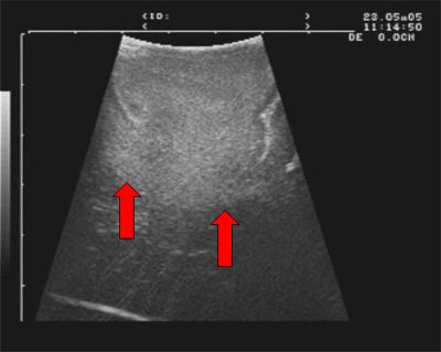
Fig.3.9: Intra-operative echogram showing the white round image of a large low-grade
glioma. This is the same case as in Fig. 3.5


 Computerized Axial Tomography
 Previous Page
