


 Computerized Axial Tomography
TOOLS FOR DIAGNOSTIC IMAGING OF BRAIN TUMORS
Traditional X-rays are useless for the diagnosis of intracranial tumors, except for calcified lesions.
In the last 30 years, thanks to the introduction of computers, less invasive diagnostic imaging tools have been developed.Computerized Axial Tomography (CAT Scan or CT Scan) and Magnetic Resonance Imaging (MRI) use computerized methods to create a direct image of the brain. Contrast enhancement may depict the lesions more clearly from the surrounding brain. This effect is due to the abnormal vascular circulation in and around the tumor (the blood-brain barrier is disrupted). CT and MRI are complementary. MRI is more sensitive for detecting small lesions and displaying anatomical details. CT is more sensitive for calcified lesions and for tumors that destroy the bone: skull or spine.
CT scan is a technique which is based on a complex X-ray machine which performs thousands of circular scans. It is equipped with a computer, which converts the signals into a digital map and then into images. The use of a iodinated contrast medium is mandatory for the diagnosis of brain tumors. CT scan without contrast is used in emergency care and traumatology. It is fast and cheap. It is very useful for studying bone and surrounding tissues: skull and spine. With modern software and hardware, CT scan is useful to reconstruct the images of the arteries and veins of the brain (Fig. 3.1).
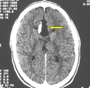
Fig. 3.1: CT scan of a corpus callosum lipoma. This fatty tumor appears as black.
This is a tunnel-shaped machine. It is based on different physical principles: it does not use X-rays. In the machine, a magnetic field is activated and exposed to radio frequency (RF) pulses. The magnetic field changes the direction of the atoms of the structure being investigated. RF causes a second change. When RF is interrupted, the atoms relax and go back to their initial orientation. In this stage, depending on the tissue being investigated, the atoms release a variable amount of energy. These energy signals are fed to a computer, which builds a digital map. From the map, the computer reconstructs the images that we see. MRI gives direct images along planes in space. CT scan gives direct images on the axial plane: the other images (lateral, coronal and oblique views) are reconstructed by the computer.
The contrast medium used in MRI is non-ionic and different from the one used in CT.
At present, MRI is the most important tool for the diagnosis of brain tumors and spinal tumors.
People with pace-makers or magnetic metal prostheses are not allowed to enter the MRI room.
Claustrophobic people do not tolerate this examination.
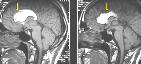
Fig.3.2: MRI of the same patient as in Fig. 3.1. Fat appears white on the MRI image.
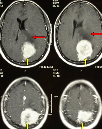
Fig. 3.3: MRI of a large left parietal meningioma. This is a benign brain tumor that may reach substantial volumes.
Page 6
 Previous Page
