


 Introduction
Glia
The glia is the supportive tissue of the brain. Its cells permit nutrition and connections to develop and exist. The most common glial cells are the astrocytes and the oligodendrocytes. Unlike neurons, glial cells are capable of reproduction. The ependymal cells are those which form the surface of the cerebral cavities, called ventricles, and are a kind of glial cells. Most of the primary cerebral tumors originate from these cells: gliomas, which may grossly be subdivided into astrocytomas, oligodendrogliomas, glioblastomas and ependymomas.
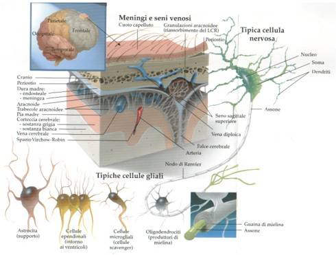
Fig. 1.5: This figure shows a section of the brain and its meningeal layers. From the outside to the inside: skin of the head, skull, meningeal layers & dura, arachnoid, pia, cortex, grey matter and white matter.
They are not nervous but connective tissue. They cover the entire CNS. The most external layer, the dura mater, is fibrous and very resistant. Under the dura, there is the arachnoid. The pia is firmly attached to the surface of the brain or spinal cord. Between the arachnoid and pia, there is the CSF. In the subarachnoid space, there are arteries and veins before their divisions and perforation of the pia to enter the brain tissue. A large layer of dura divides the cerebral hemispheres from the cerebellum and brainstem: the tentorium. This is a curtain with a large hole that permits the passage of the descending structures. It divides the skull in the supratentorial and infratentorial compartments.
A very frequent, generally benign, intracranial tumor arises from the meninges: the meningioma.
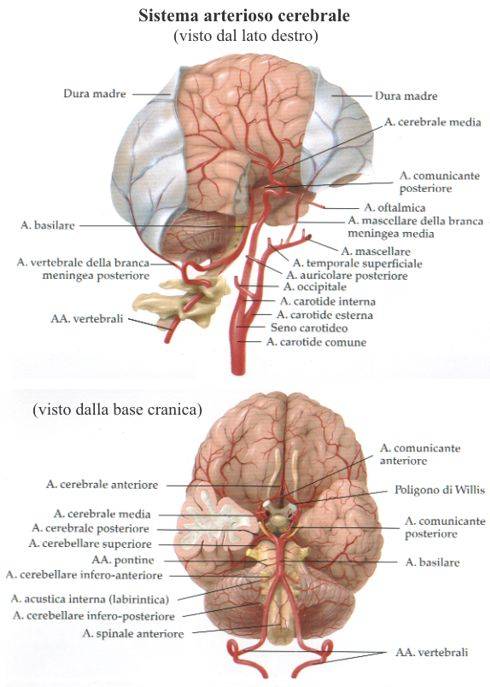
Fig. 1.6: This picture shows the arteries of the brain, seen from below and laterally. As is obvious, a tumor arising at the base of the brain, i.e. a location with big arteries, will be technically more challenging than one arising from the surface of the hemisphere.
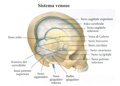
Fig. 1.7: This one shows the large dural sinuses, where the venous blood exits from the intracranial structures. The cerebral veins flow into the sinuses.
The neurons are the "thinking" cells of the brain. They are connected with the overall brain and cerebellum by axons and dendrites. They are extremely specialized: motor, sensory, visual and so on. Actually, they constitute the most complex being of the universe.
Pineal Gland
This is a small structure located under the posterior part of the corpus callosum. It produces melatonin. Its role in humans is still unclear. It is likely to play a role in the control of biorhythms.
Skull
This is the bony box that contains and protects the brain. There is a cranial vault and a cranial base. On the base, there is a large hole: the occipital hole. Through this hole, the spinal cord (a prolongation of the brain) passes into the spinal canal. On the base, there are other small holes that permit the passage of nerves, arteries and veins.
Spinal Cord
It is the lowermost part of the CNS. Along the cord are the paths that carry motility and sensitivity to the entire body. In the spinal cord, the white matter is external and the grey matter is internal. The internal part of the spinal cord contains the nuclei of the peripheral nerves. The cord is located in a rigid-elastic ligamentous bone tube that is the spinal canal. In adults, the cord ends at L1. The nerves for the lower limbs, bladder, rectus and genitals arise from the most caudal part of the cord and constitute the so-called "cauda equina".
The nerves for the upper limbs arise from the cervical segments of the cord and constitute the brachial plexus. The same tumors that arise from the brain may arise from the cord: gliomas, meningiomas, neurinomas and ependymomas.
Ventricles
They are four connected cavities: the lateral ventricles (one in each hemisphere), the third ventricle and the fourth, which is located anteriorly to the cerebellum, in the brainstem. From the fourth ventricle, the CSF flows into the spinal cord subarachnoidal space.
Page 3
 Previous page
