


 Introduction
1.2 Glossary
Basal NucleiThese are large areas of neurons (grey matter) located in the deep part of the hemispheres. The largest one is the thalamus (Fig. 1.3). They are important centers for psychic and motor-sensory functions. They are connected with the cortex, cerebellum and spinal cord. A typical illness of the basal nuclei is Parkinson's disease.
Brain - cerebral hemispheres
The largest structure in the skull is the brain, which is divided in two hemispheres, right and left: the right for the left side of the body and the left for the right one. In right-handed people, the left is the dominant hemisphere. In left-handed people, it is the other way around. The dominant half of the brain generally accommodates the areas of superior psychic activity: speaking, reading, ability to perform calculations and writing. Each hemisphere is divided in four parts, called cerebral lobes: frontal, parietal, temporal and occipital.
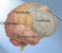
Fig. 1.1: Lobes of the right brain hemisphere.
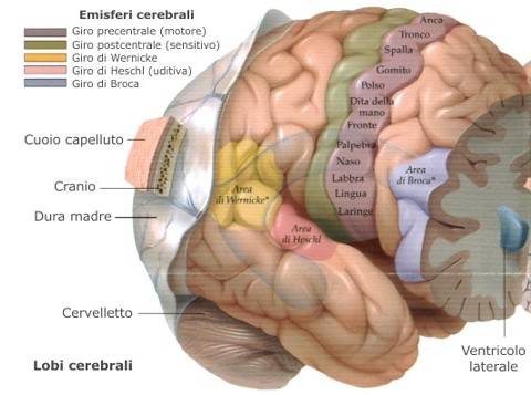
Fig. 1.2: External surface of the brain, with lobi and related functional areas: motor cortex and speaking areas.

Fig. 1.3: Internal surface of a brain hemisphere where the corpus callosum and fornix are shown: these are key structures that link the two hemispheres. The thalamus is an important sensory and psychic nucleus. Note also the pituitary gland, the cerebellum and the cranial nerves:
- Olfactory nerve: smell and taste
- Optic nerve: vision
- Oculomotor nerve: ocular movements
- Throclear nerve: ocular movements
- Trigeminal nerve (sensory nerve for the face and motor nerve for chewing)
- Abducens nerve: ocular movements
- Facial nerve (motor nerve for the face )
- Auditory and vestibular nerve : hearing and balance
- Glossopharyngeal nerve: swallowing
- Vagus nerve (gastrointestinal tract, heart)
- Accessory nerve (muscles of the neck and shoulder)
- Hypoglossal nerve (tongue)
It is the part that connects the brain to the spinal cord. In the brainstem, there are the vital nuclei for breathing and cardiac activity. It also contains the nuclei of the cranial nerves. Anatomically, it is divided in three portions: mesencephalon (the most cranial), then the pons and the bulb (the most caudal). A complex neuronal network, the reticular substance, is located in the brainstem. This is connected with the overall brain and governs our consciousness.
Any important lesion of the brainstem is lethal.
Cerebellum
This is the second largest structure of the brain. It is formed of two hemispheres and a median portion, called vermis. It is attached to the brainstem through the cerebellar peduncles. It is located in the posterior cranial fossa, under the tentorium. Its functions are important for coordination, balance and gait. It is connected with the spinal cord and the cerebral hemispheres.
Cerebrospinal Fluid (CSF)
The brain and the spinal cord float in a watery pillow: the cerebrospinal fluid (CSF), which is produced in the cerebral ventricles and resorbed in the subarachnoidal space. The CSF changes continuously. A lumbar puncture is used to collect the CSF for examination. The CSF pathways may be obstructed by tumors or be defective in their functioning. In these cases, the CSF builds up in the ventricles, causing hydrocephalus and increase of intracranial pressure.
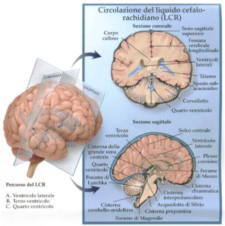
Fig. 1.4: Pathway of the CSF from the ventricles to the basal cisterns of the subarachnoid space. Building up of the CSF in the ventricles causes hydrocephalus.
It is a very important structure, as it connects the two hemispheres. Extended lesions of this connection cause severe psychic disorders.
Hypophysis or Pituitary Gland
This is an endocrine gland, large as a bone and located at the base of the brain, under a small dural curtain. It is linked to the hypothalamus by the pituitary stalk. It is located in a bony pocket, called sella turcica or sellar cavity. The pituitary is an interpreter of the hypothalamus language for the other glands of the body. Without the pituitary, thyroid, sexual glands and adrenal glands would not be able to understand the orders coming from the brain and mediated by the hypothalamus. The pituitary also produces the growth hormone, which has a direct action. The posterior part of the pituitary is formed of nervous tissue and releases vasopressin (ADH - regulating water metabolism) and oxytocyn (regulating delivery). Tumors originating from the pituitary are rather frequent, benign and defined as pituitary adenomas.
Hypothalamus
This is the vital structure that governs vegetative and sexual life. It is located anteriorly to and around the third ventricle. It regulates metabolism and reproduction. It is connected with the overall brain in order to adjust metabolism to external stimuli, needs and emotions.
Page 2
 Previous Page
