Other sites of occurrence are very rare.
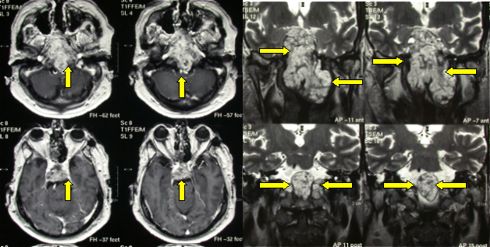
Chordomas of the cranial base present with cranial nerve deficits. Both CT scan and MRI show extensive bone destruction and adjacent soft tissue involvement.

Fig.5.48: Large cranial base chordoma in axial (left, yellow arrows) and frontal views (right).

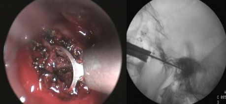
Fig.5.49: Intra-operative endoscopic view of the same case. On the right, radioscopy through a transnasal approach.
Spinal chordomas are located at the sacrum. They may be removed by very experienced spinal surgeons, sometimes in cooperation with abdominal surgeons.
Chondromas and Chondrosarcomas are rare. In the skull, they may recall meningiomas. Chondroma is a benign cartilaginous tumor, located at the cranial base, sometimes extended to the orbit. It generates from the basicranial synchondrosis. Chondrosarcoma is the malignant type of chondroma. Clinically, the latter tumor behaves like a chordoma.
Osteoma is a benign bony tumor that presents as a hard skull swelling. It is visible at plain radiographs; details are seen at CT scan with bone window. Sometimes, skull osteoma is the external expression of an underlying meningioma. Osteomas may be removed owing to cosmetic deformation of the skull, particularly if they are located in the fronto-orbital region, or owing to patient's pain and distress. The removed bone must be replaced. Use may be made of several materials: porous ceramics, titanium, acrylic. Whatever the material used, custom-made cranioplasty should be performed in fronto-orbital locations. In other sites, cranioplasty is simpler, since the cosmetic result is less important.
Page 23


 Glioma
 Previous Page