This technique produces images in a faster sequence, thus detecting brain areas with higher oxygen consumption, i.e. those activated upon the examination. During functional MRI, a person may talk and activate his/her language areas. This type of MRI identifies these eloquent areas. The same occurs if the person being examined is invited to move a finger or a foot, think of colors or imagine a scene. Functional MRI is very useful in planning neurosurgery in the parts of the brain that are "critical" or "eloquent".
The data from this investigation are fed to and correlated with those provided by the
neuronavigator upon surgery.
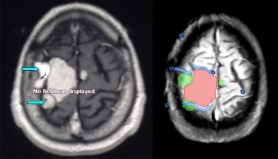
Fig.3.4: Functional MRI of a meningioma located on the motor cortex. On the left, the tumor (the large white area) with adjacent areas activated by movements. On the right, the reconstruction for the
neuronavigator. The tumor is pink, the motor cortex is green, the important veins are blue.
This examination is based on the capability of the scanner to identify the components of the proteins contained in a given area of the brain and/or brain tumor.
The occurrence of and relationships between the various amino acids may differentiate the various tissues: brain tumor, brain edema, radionecrosis and other diseases which may be mistaken for tumors upon standard MRI.
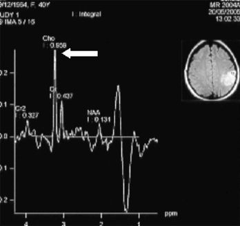
Fig. 3.5: Spectroscopic MRI of a left parietal low-grade glioma. The examination detects amino acids that are more present in tumoral tissue than in other diseases.
This is a particular type of MRI which investigates the blood flow rate. It may thus differentiate between arteries (where the flow rate is faster) and veins (where the flow is slow). The reconstructed image is a static one. It is very helpful in planning the surgical procedure and may be correlated with other data, which are processed by the
neuronavigator.
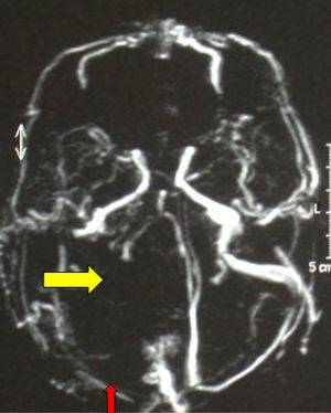
Fig.3.6: Angiographic MRI scan: the left half of the image clearly shows few vessels, i.e. the site of a large meningioma. The venous sinuses are already occluded by the tumor.
This is a new technique, which has been made possible by modern and more powerful MRI machines. With this technique, we may visualize not only the functional areas of the brain cortex in which we are interested, but also the tracts departing from them and trace their pathways in the brain.
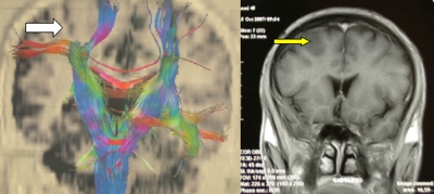
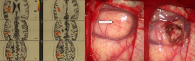
Fig. 3.7: The upper left image is a tractographic one. The white arrow points to the tumor that splits the motor tracts; on the other side, the same tracts are well visible. In the upper right image, the yellow arrow points to a low-grade glioma as seen in conventional MRI. Bottom left: the functional motor areas are orange. Bottom right: operative views of the tumor before and after
neuronavigator-guided removal.


 Computerized Axial Tomography
 Previous Page
